Retinal detachment is a condition in which the light-sensitive retinal layer lining the back wall of the eye separates from the underlying supportive tissue. Retinal detachment (the "separation of the retina" that develops after a retinal tear or for other reasons) is a serious eye condition that requires urgent intervention. If left untreated, it can lead to permanent vision loss. In this guide, you will find a plain-language explanation of what retinal detachment is, its types, warning signs, and diagnostic and treatment approaches.
What is retinal detachment, and why is the retina important?
Retinal detachment is the separation of the retina from the inner surface of the eyeball, causing it to lose its blood supply and function. The retina is the tissue that produces visual signals; once it detaches, its cells cannot receive enough oxygen and nutrients.
In brief:
- Retinal detachment can occur without pain, so it can easily go unnoticed.
- Timely treatment is decisive for the visual outcome.
- When symptoms are noticed, you should see an eye specialist without delay.
What is the structure and function of the retina?
The retina is a thin, transparent layer of nerve tissue located at the back of the eye. It works like the film or sensor in a camera: it converts the light entering the eye into electrical signals and transmits them to the brain via the optic nerve.
- Macula: Located at the center of the retina; it is essential for reading, recognizing faces and seeing fine detail.
- Peripheral retina: Provides side vision and night vision.
- Retinal pigment epithelium and choroid: The underlying layers that nourish and support the retina.
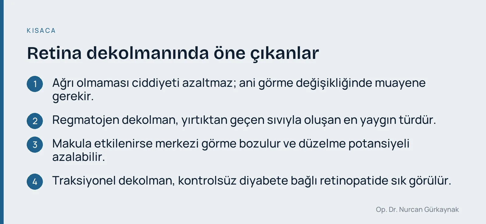
In retinal detachment, the separation can begin at the periphery and progress toward the macula. When the macula is also affected, central vision is impaired and the potential for recovery may decrease. For other retinal conditions, you can visit our retinal diseases page.
What are the types of retinal detachment?
Retinal detachment is divided into three main types according to its mechanism. Identifying the type directly influences the choice of treatment.
| Type | Basic mechanism | Commonly seen in |
|---|---|---|
| Rhegmatogenous | A tear or hole forms in the retina, and fluid passes through the opening beneath the retina | Advanced age, high myopia, trauma, after cataract surgery |
| Tractional | Scar tissue pulls on the retina and separates it | Diabetic retinopathy, vascular occlusions, some inflammatory conditions |
| Exudative (serous) | Fluid accumulates beneath the retina; there is no tear | Inflammatory diseases, tumors, fluid leakage from blood vessels |
What is rhegmatogenous retinal detachment?
Rhegmatogenous retinal detachment is the most common type of retinal detachment, in which fluid passing through a tear or hole in the retina separates it from the underlying tissue. It takes its name from the Greek word "rhegma" (tear). It most often occurs when the vitreous gel inside the eye shrinks with age and pulls on the retina.
What is tractional retinal detachment?
Tractional detachment occurs when fibrovascular (scar-like) tissue on the surface of the retina pulls on it. It is most commonly seen in proliferative retinopathy caused by uncontrolled diabetes.
What is exudative (serous) retinal detachment?
In exudative detachment, there is no tear in the retina; instead, fluid accumulates beneath it. Treatment is directed at the underlying cause.
What are the causes and risk factors of retinal detachment?
Retinal detachment is not caused by a single factor; many factors related to age, eye structure and accompanying diseases play a role. The main risk factors are:
- High myopia: When the eyeball elongates, the retina thins and becomes prone to tearing.
- Advanced age: Changes caused by aging of the vitreous gel (posterior vitreous detachment) increase the risk.
- Eye trauma: Blunt or penetrating injuries can cause tears in the retina.
- Previous eye surgery: The risk may increase slightly after cataract surgery.
- A history of retinal detachment in the family or in the other eye.
- Peripheral degenerations of the retina (for example, thinned areas known as "lattice").
- Diabetes and other vascular diseases: Particularly associated with tractional detachment.
- Inflammatory eye diseases.
Having risk factors does not mean you will develop a detachment; it simply underlines the importance of regular check-ups.
What are the symptoms of retinal detachment?
The typical symptoms of retinal detachment are sudden vision changes that are often painless. The main signs are:
- Floaters: A sudden increase in black dots, web-like spots or fly-like shapes in front of the eye.
- Flashes of light (photopsia): Lightning-like flashes, especially seen in the dark or with eye movement.
- Curtain or shadow: A sensation of a dark curtain that starts at one edge of the visual field and moves toward the center.
- Blurred vision and straight lines appearing curved.
- Reduced central vision (when the macula is affected).
Is retinal detachment painful?
No, the retinal tissue has no nerve endings that sense pain; therefore, retinal detachment is usually painless. The absence of pain does not make the condition any less serious. If you experience a sudden change in vision, you need an examination even if there is no pain.
When should you seek emergency care?
If any of the following occurs, see an eye specialist or go to the nearest emergency department without delay:
- A sudden appearance of many new floaters in front of the eye
- Recurrent flashes of light
- A shadow, curtain or darkening in your field of vision
- A sudden change in vision after an eye injury
- Any new symptom in people with high myopia or a previous retinal detachment
A brief, isolated eyelid twitch is usually not related to the retina; for more on this, see our article what causes eye twitching and when to see a doctor. However, if there is a curtain or flashes of light in your field of vision, the situation is different and needs to be evaluated.

How is retinal detachment diagnosed?
Diagnosis is based on a detailed eye examination. The following methods are commonly used:
- Dilated fundus examination: The pupil is dilated with eye drops, and the retina is examined with special lenses and light systems. Tears, holes and the area of detachment can be seen this way.
- OCT (optical coherence tomography): A contactless, painless imaging method that shows the layers of the retina in cross-section. It is particularly useful for assessing the condition of the macula.
- Ocular ultrasound (B-scan): Helps evaluate the state of the retina when the inside of the eye cannot be seen because of bleeding or cloudiness.
- Visual acuity and visual field tests.
The examining physician decides which tests are necessary.
What happens if retinal detachment is left untreated, and how is it treated?
Untreated retinal detachment can progress and lead to permanent vision loss in the affected eye. The longer the retina remains detached, the greater the cell damage. For this reason, treatment is surgical as a rule, and timing is important. The detachment cannot be expected to resolve on its own with medication or eye drops.
The approach is planned according to the type and extent of the detachment and the condition of the macula. The main methods are:
- Laser photocoagulation or cryopexy (freezing): Applied around tears that have not yet progressed to detachment, "welding" them to prevent fluid from passing through.
- Pneumatic retinopexy: Based on injecting a gas bubble into the eye to push the retina back into place; it can be used in selected patients.
- Scleral buckle: A silicone band is placed on the outside of the eyeball to push the wall inward.
- Vitrectomy: An intraocular surgery in which the vitreous gel is removed and the retina is repositioned, supported with gas or silicone oil if necessary. This method is often required in tractional detachment.
In exudative detachment, the priority is treating the underlying disease that causes the fluid buildup. The appropriate method is decided after a personalized examination; results vary depending on the patient and the characteristics of the disease, and no definite guarantee can be given.
What does the post-surgery process and recovery look like?
The post-operative process differs depending on the method used. In general, the following points are important:
- Drops: Eye drops that prevent infection and reduce inflammation are used as recommended by your doctor.
- Positioning: If a gas bubble was used, your doctor may ask you to maintain a specific head position.
- Travel and altitude: While a gas bubble is present, air travel and high altitude can change eye pressure dangerously; always consult your doctor.
- Activity restrictions: You may be asked to avoid heavy lifting and sudden movements for a certain period.
- Follow-up: Scheduled check-ups should not be missed; vision usually improves gradually over weeks to months.
If you experience increased pain, a new decrease in vision, redness or a new sense of a curtain, contact your doctor without waiting.
How can you protect yourself from retinal detachment?
Not every retinal detachment can be prevented, but the risk can be reduced and the chance of early diagnosis increased:
- People with high myopia, a family history of detachment or previous eye surgery should have regular dilated fundus check-ups.
- Conditions such as diabetes and hypertension should be kept under control.
- Protective eyewear should be used in high-risk sports and jobs.
- If sudden floaters or flashes of light occur, get examined without delay.
- Other eye conditions such as glaucoma should also be monitored; for details, see our glaucoma (eye pressure) page.
Frequently asked questions
Does retinal detachment heal on its own?
No. A true retinal detachment does not resolve on its own and usually requires surgery.
Does retinal detachment always lead to permanent vision loss?
Not always. When treated early and before the macula is affected, the likelihood of preserving vision is higher. The risk of permanent loss increases with delay. Individual results depend on your doctor's evaluation.
Does seeing floaters mean retinal detachment?
No. Many floaters are harmless and age-related. However, floaters that are new, increasing in number or accompanied by flashes of light always require an examination.
Can retinal detachment occur in both eyes?
Yes. In people who have had a detachment in one eye, the risk is higher in the other eye, so the other eye is also monitored regularly.
Where can you go for retina health?
Eye examinations and evaluations for retinal diseases are carried out at the practice of Op. Dr. Nurcan Gürkaynak in Alsancak, İzmir; for detailed information, you can visit our retinal diseases page. This article is for general information only; for diagnosis and treatment, you should always consult an eye specialist.
Frequently Asked Questions
What is retinal detachment?
Retinal detachment is a condition in which the retina, the layer of tissue at the back of the eye, separates from the tissue beneath it. It is a serious condition that can lead to vision loss.
What are the types of retinal detachment?
There are three main types of retinal detachment: rhegmatogenous, tractional, and exudative. Each has different causes, and treatment methods vary accordingly.
What are the symptoms of retinal detachment?
Symptoms of retinal detachment include sudden vision loss, flashes of light, floaters, and a shadow or dark curtain in the field of vision. These symptoms require emergency medical attention.
How is retinal detachment diagnosed?
Retinal detachment is diagnosed through an eye examination and a dilated fundus examination. Your eye doctor uses specialized instruments to assess the condition of the retina.
What are the treatment options for retinal detachment?
Treatment of retinal detachment usually requires surgery. Methods such as laser therapy, vitrectomy, or scleral buckling may be used.
Is it possible to prevent retinal detachment?
To help prevent retinal detachment, it is important to have regular eye examinations and avoid eye injuries. People with risk factors in particular should be extra careful.